Radiation Therapy

IsoPhantom: A Customizable 3-D Phantom for Improved Radiation Dosimetry
WARF: P100285US01
Inventors: Benjamin Nelms
The Wisconsin Alumni Research Foundation (WARF) is seeking commercial partners interested in developing a radiation dosimetry quality assurance system to improve the safety and quality of radiation delivery.
Overview
Radiation therapy methods such as external beam radiation therapy are administered by directing beams of radiation toward a defined target volume in a patient. A radiation therapy plan is used to determine how much radiation to deliver and from what locations while ensuring that healthy tissue surrounding the target tissue does not receive excessive radiation. Modern radiation therapy methods such as intensity modulated radiation therapy (IMRT) and volume-modulated arc therapy (VMAT), coupled with image-guided radiation therapy (IGRT), allow for highly customized dose optimization. However, there is still potential for the patient to receive undesired radiation doses, i.e., it is possible for the actual delivered dose to be different than the planned dose, and such errors can have a negative clinical impact.
The process of pretreatment verification of per-patient radiation dose commonly is called dose quality assurance (dose QA). Sophisticated radiation therapy modalities featuring rotational therapy use a 3-D phantom for dose QA instead of the common 2-D arrays used for static gantry angle IMRT. However, 3-D phantoms generally are coupled with either 2-D detection arrays or 3-D detection systems. The use of 2-D detection arrays presents problems of angular dependence and may only measure dose along a single plane, which limits the ability to detect errors. 3-D dosimetry arrays are expensive and may have limited (i.e., fixed, regardless of plan type) or unsuitable detector placement. A customizable system for per-patient radiation dose QA would be desirable to provide greater flexibility in radiation dosimetry without requiring the purchase of an expensive new dosimetry system.
The process of pretreatment verification of per-patient radiation dose commonly is called dose quality assurance (dose QA). Sophisticated radiation therapy modalities featuring rotational therapy use a 3-D phantom for dose QA instead of the common 2-D arrays used for static gantry angle IMRT. However, 3-D phantoms generally are coupled with either 2-D detection arrays or 3-D detection systems. The use of 2-D detection arrays presents problems of angular dependence and may only measure dose along a single plane, which limits the ability to detect errors. 3-D dosimetry arrays are expensive and may have limited (i.e., fixed, regardless of plan type) or unsuitable detector placement. A customizable system for per-patient radiation dose QA would be desirable to provide greater flexibility in radiation dosimetry without requiring the purchase of an expensive new dosimetry system.
The Invention
A UW–Madison researcher has developed a system and method for utilizing available detector arrays with a highly configurable 3-D phantom to acquire accurate 3-D dosimetry information. This radiation dosimetry quality assurance system is configured to measure actual radiation dose delivered by the radiation delivery system during a planned medical procedure.
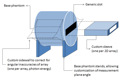
The system consists of a 3-D phantom extending along a longitudinal axis to form an exterior surface and an interior volume, a passage that extends the interior volume of the phantom to receive a detector array, an angular-compensation system coupled to the surface of the 3-D phantom to control any angular dependence of the detector array during dose measurement and a detector-angle adjustment system to allow positioning of the detector array with respect to the radiation source (see the figure below for a general schematic of the system). The system: 1) corrects for device-dependent angular dependence, 2) optimizes the measurement plane(s) to any plane rotated about the longitudinal axis to maximize sensitivity specific to each radiation plan, and 3) is capable of serving all 2-D dosimetry arrays. The system can be utilized with flexible analysis software to integrate 2-D array measurements into a 3-D analysis engine and graphical user interface.
The system consists of a 3-D phantom extending along a longitudinal axis to form an exterior surface and an interior volume, a passage that extends the interior volume of the phantom to receive a detector array, an angular-compensation system coupled to the surface of the 3-D phantom to control any angular dependence of the detector array during dose measurement and a detector-angle adjustment system to allow positioning of the detector array with respect to the radiation source (see the figure below for a general schematic of the system). The system: 1) corrects for device-dependent angular dependence, 2) optimizes the measurement plane(s) to any plane rotated about the longitudinal axis to maximize sensitivity specific to each radiation plan, and 3) is capable of serving all 2-D dosimetry arrays. The system can be utilized with flexible analysis software to integrate 2-D array measurements into a 3-D analysis engine and graphical user interface.
Applications
- Customized radiation therapy treatment planning and dose verification
Key Benefits
- Offers flexible dose measurement through critical targets
- Improves positioning relative to the patient for dose verification
- Reduces cost of treatment by offering a less expensive alternative to previously developed 3-D dosimetry phantoms
- Allows use of existing 2-D arrays inside a customizable 3-D phantom
- Enables use with existing 2-D radiation therapy detector software
Tech Fields
For current licensing status, please contact Jeanine Burmania at [javascript protected email address] or 608-960-9846
Figures
